By Jane Metlikovec
Dr Rishi Ramani won first prize for his poster on the creation of a deep learning model for accurate detection of graded oral dysplasia in a carcinogenic murine model, highlighting a novel pipeline for oral cancer prevention at the 2nd Global Oral Cancer Forum (GOCF 2024) in Kuala Lumpur, Malaysia.
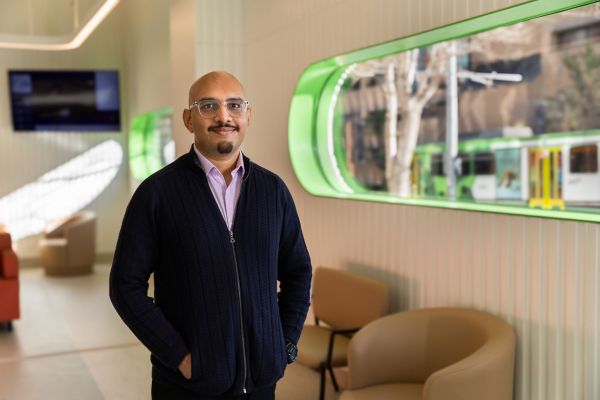
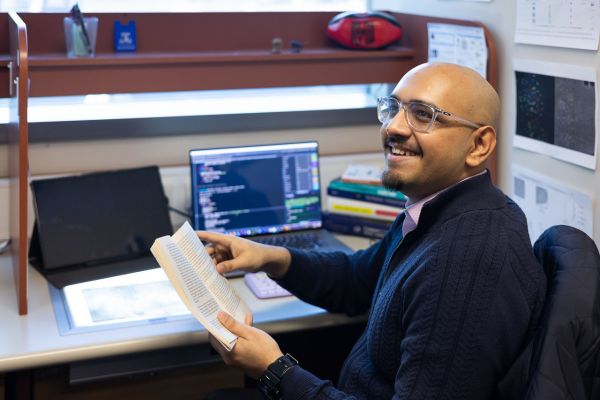
A Python-programming dentist may seem unusual, but an unwavering dedication to improving oral cancer detection sometimes means PhD candidate Dr Rishi Ramani is more likely to be found coding Artificial Intelligence (AI) algorithms than examining dental records.
The award-winning researcher behind new ‘digital biopsies’ applied to join the World Health Organization’s ‘Global Initiative on AI 4 Health’ to help develop ethical standards for the use of AI in dentistry, but he immediately brings his ground-breaking innovative work back to patients.
“Say you’re a patient, and you’ve noticed something is a little different in your mouth,” Dr Ramani says.
“And so, you wait for a week or two, then you go to a dentist. The dentist might then decide you need to go to a specialist, but there are only 50 of those specialists in the entire country. So, you wait for, say, six months. That is many months of stress and anxiety, and in that time, it might be nothing, or you might have a condition that’s progressing.”
Credit: Peter Casamento
Imagine if that first dentist you see could do a simple non-invasive image-based screening, right there in the chair, and put those images straight into an AI system that can provide an accurate diagnostic result immediately, right then and there. Dr Rishi Ramani
That’s the goal that Dr Ramani - who trained as a dentist earlier in his native India - has been working towards through his PhD at Melbourne Dental School, with his work recently published in Nature Portfolio, and winning the Global Oral Cancer Forum poster competition in Kuala Lumpur, 2024.
A bold new vision for preventative health
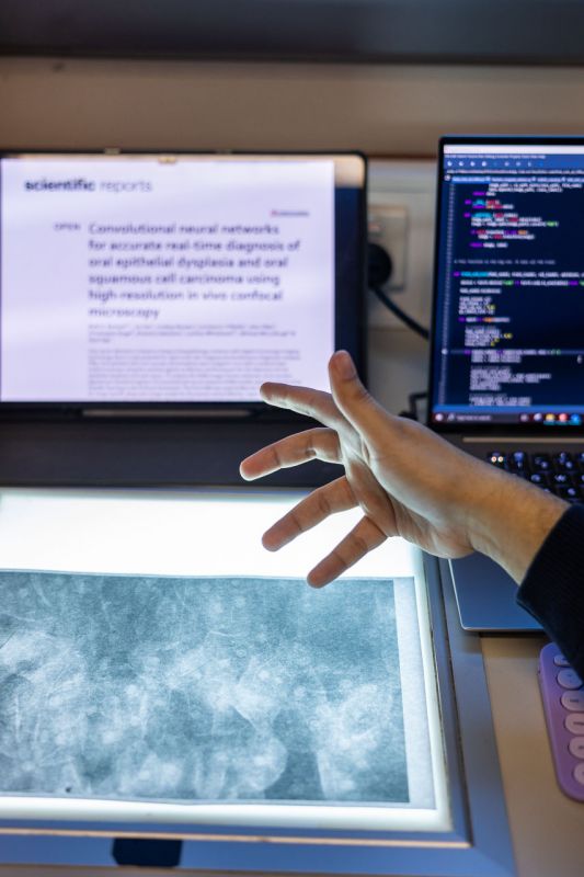
As Dr Ramani explains, the concept works like this: A patient visits a dentist, who presses a small probe against a suspicious patch in their mouth. The probe makes detailed images that are different from an X-ray; they can reveal living cells and nuclei in soft tissues, like the cheek.
These images are then fed into an AI system, which has learned how to identify cancer images from being trained on similar images. It understands patterns in the image that might correspond to signs of cancer. As Dr Ramani says, “Imagine the impact of being able to get instant results for something so potentially life-changing, in a completely painless way.”
It’s an exciting prospect, but to develop the idea, Dr Ramani had to master the latest technology – not to mention develop a brand-new AI system of his own. “We couldn’t just use off-the-shelf AI, because these new kinds of cellular images are completely novel and nobody has trained a machine to recognise oral cancer in this specific way before.”
So, Dr Ramani sat down to learn Python and PyTorch, some of the programming languages powering today’s AI tools, like ChatGPT.
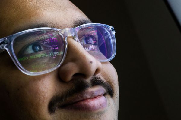
![The silhouette of PhD student, Dr Rishi Ramani with a digital display showing code in the background.]](https://dental.unimelb.edu.au/__data/assets/image/0006/5414433/varieties/medium.jpg)
Creating a new AI system from scratch
It was a learning curve, but Dr Ramani relished the challenge and built his new AI system. What wasn’t so enjoyable, he says, was the weeks both Dr Ramani and his supervisors spent manually circling nuclei on image upon image to feed into the system.
We had over a thousand images that we needed to label manually so I could build this segmentation and classification model that would automate this identification process. Dr Rishi Ramani
“It was very difficult, and you need really high-quality images to get it right and for it to deliver value. It’s like that old saying, ‘garbage in, garbage out’, so it was really important that what we put into the system was going to be of the highest quality so that it could be clinically useful.”
Where AI shines - and where there’s work to be done
To date, Dr Ramani’s program has revealed both some exciting results and areas for improvement.
The first AI model worked well at filtering out poor-quality images, Dr Ramani says, meaning blurred or defective images wouldn’t be considered for patient results. When it came to a particular type of image, where the fluorescent dye acriflavine was used, Dr Ramani’s second AI diagnostic model correctly identified all the high-grade dysplasia (which shows significant abnormalities at high risk of cancer), during his testing.
Speed, too, was a clear stand-out, with each new patient image classified in 0.03 seconds.
Data scarcity was an issue, however, with only about 20 per cent of the images captured being of good quality. As Dr Ramani says, “this shows imaging inside someone’s mouth is quite challenging with the patient moving and the saliva that can get in the way.”
But working at this new frontier has now propelled Dr Ramani into an exciting new professional global community; he’s now part of the World Health Organisation’s Global Initiative on AI for Health.
“We are a group of researchers from around the world working to create standards and guidelines, essentially establishing how AI should be used in dentistry ethically and objectively, which is so important and really exciting," he says.
Read more about Dr Ramani’s involvement in the World Health Organization's Global Health Initiative on AI for Health.
Credit: Peter Casamento